
Mohs Surgery is associated with the highest possible cure rate
Mohs Micrographic Surgery is a highly specialised and precise surgical technique used to treat skin cancers. It combines surgical excision with complete microscopic margin analysis, ensuring the highest possible cure rate (up to 99%) while preserving healthy tissue.
International skin cancer guidelines recommend Mohs Surgery as first line treatment and gold standard for skin cancers at high risk for recurrence and in cosmetically sensitive and/or functionally important areas.
It has been proven to provide to best chance of cure, being the most cost-effective and associated with best cosmetic outcomes when treating skin cancer.
What makes Mohs Surgery the best choice for skin cancer treatment?
Unmatched Precision
Unlike traditional surgery, Mohs Surgery removes the cancerous tissue layer by layer, examining 100% of the margins to ensure complete removal.
Minimised Tissue Loss
Only cancerous tissue is removed, preserving as much healthy skin as possible – potentially leading to simpler reconstruction and better cosmetic outcomes.
High Cure Rates
Offers the highest cure rate, making it the most effective method for skin cancer treatment.
The Mohs procedure explained
Mohs surgery is performed under local anaesthetic in a specialised, sterile environment. Your Mohs surgeon will begin by removing the visible tumour and a thin layer of surrounding tissue. This tissue is immediately processed in an on-site Mohs lab. The Mohs specific method of processing allows tissue slides to be produced that show the whole, 100% complete cut surface around and underneath the tumour. These slides are then examined by the Mohs surgeon and will show very accurately any remaining tumour.
If any cancer cells are found, further excision of the affected area is performed. This continues layer by layer until all cancer is gone, ensuring that as much healthy tissue as possible is preserved. Once the cancer is completely removed, the wound is carefully repaired to ensure the best cosmetic outcome and healing.
The Mohs Surgery Process

Step 1: Skin cancers often extend beyond their visible borders, and these extensions (roots) can cause the tumour to recur if not completely removed.
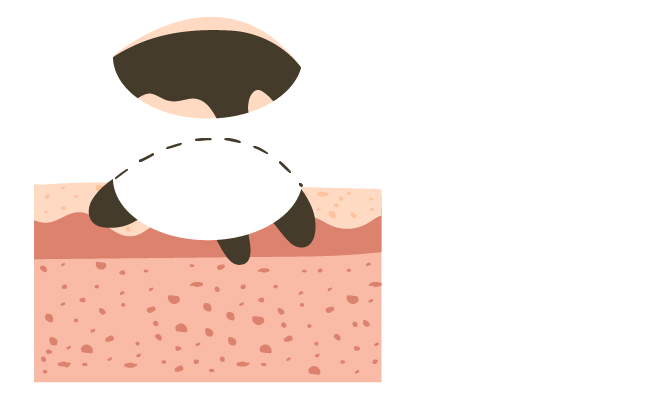
Step 2: The Mohs technique begins by removing the visible tumor with thin layer of healthy tissue.
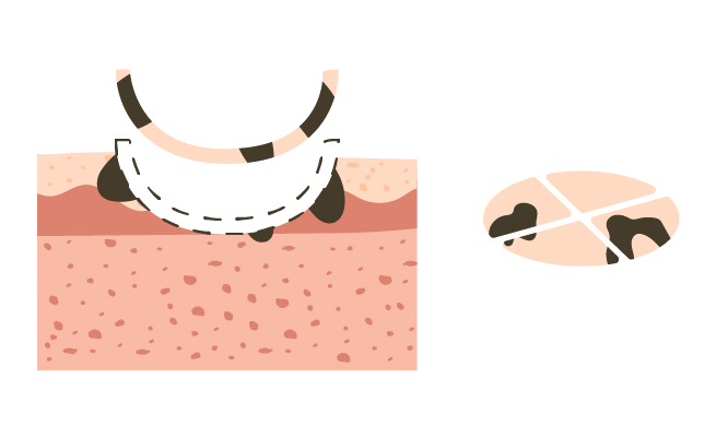
Step 3: This tissue is mapped and examined under a microscope by the surgeon to check for any cancerous cells.
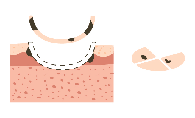
Step 4: If cancerous tissue remains, additional thin layers are removed, precisely focusing on the areas where the cancer persists, until the tumor is entirely eliminated.

Step 5: Mohs surgery is unique in that it ensures the entire tumor is removed while preserving the maximum amount of healthy tissue. This minimizes scarring and reduces the risk of cancer recurrence.
Traditional Wide Local Excision
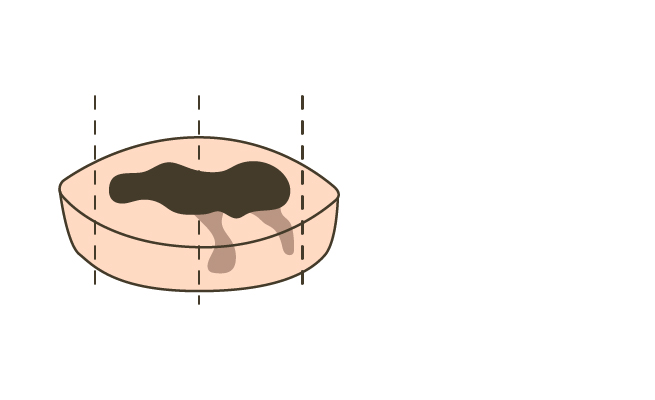
Step 1: A wide elliptical excision is made around the tumour (5mm to 10mm).
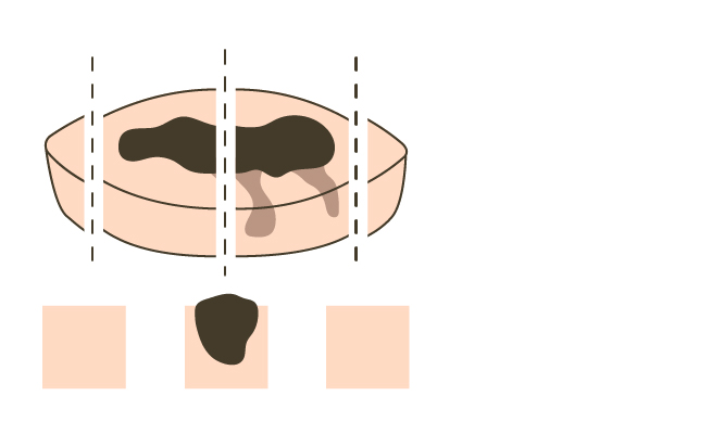
Step 2: During traditional wide local excision, random vertical sectioning (often referred to as ‘breadloaf’ sections) is used for histologic examination. However, only a small fraction of the surgical margin (typically about 1%) is analysed. This limited sampling can miss extensions (roots) of the tumour that are present beyond the sampled tissue, leading to false-negative results in the histopathology. This increases the risk of recurrence due to incomplete removal of cancerous cells.
Why choose Mohs Surgery over traditional skin cancer surgery?
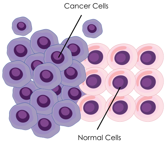
In traditional skin cancer surgery, the tumour is excised with a wide margin of healthy tissue – typically greater than 5mm for high-risk basal cell carcinomas (BCC) and greater than 10mm for squamous cell carcinomas (SCC). While this approach aims to ensure complete removal, it often results in larger surgical defects and more complex reconstructions. Additionally, the technique is somewhat “blind,” as there is no immediate confirmation that all cancerous tissue has been removed, meaning tumour cells can remain, even after sacrificing a significant amount of healthy tissue.
Histological assessment following standard excision typically involves random vertical sectioning, or “bread loafing,” where only a small portion (around 1%) of the surgical margin is examined. In contrast, Mohs surgery evaluates the entire margin (100%), offering a far more thorough assessment.
As a result, the limited sampling in traditional excision increases the risk of missing residual tumour cells, leading to potential recurrence of cancer.
Indication for Mohs Surgery according to the NCCN guidelines:
Keratinocyte Carcinomas
• Tumours of any size on the head and neck, hands, feet, pretibial and anogenital area.
• Tumours ≥20 mm in diameter on trunk and extremities
• Lesions with poorly defined borders
• Recurrent lesions (Any previous treatment)
• Lesions in sites of prior radiation therapy and scars
• Perineural invasion (invasion of nerves)
• Immunocompromised patient
• Rapidly growing tumour
• Aggressive histopathologic features
Have questions?
If you’re unsure about Mohs Surgery or want more details on the procedure, recovery, or anything else, check out our comprehensive FAQ section.